


быстрая доставка ~4 недели
Размер
- 100 μL
- 200 μL
- 50 μL
DR5/TRAILR2/TNFRSF10B Rabbit mAb (A19043)
- Описание
- Характеристики
-
Overview
Product name DR5/TRAILR2/TNFRSF10B Rabbit mAb Catalog No. A19043 Host species Rabbit Purification method Affinity purification Isotype IgG CloneNo. ARC0406 Background
The protein encoded by this gene is a member of the TNF-receptor superfamily, and contains an intracellular death domain. This receptor can be activated by tumor necrosis factor-related apoptosis inducing ligand (TNFSF10/TRAIL/APO-2L), and transduces an apoptosis signal. Studies with FADD-deficient mice suggested that FADD, a death domain containing adaptor protein, is required for the apoptosis mediated by this protein. Two transcript variants encoding different isoforms and one non-coding transcript have been found for this gene.Immunogen information
Immunogen A synthetic peptide corresponding to a sequence within amino acids 341-440 of human DR5/TRAILR2/TNFRSF10B (O14763). Sequence CFDDFADLVPFDSWEPLMRKLGLMDNEIKVAKAEAAGHRDTLYTMLIKWVNKTGRDASVHTLLDALETLGERLAKQKIEDHLLSSGKFMYLEGNADSAMS Gene ID Swiss Prot Synonyms DR5; CD262; KILLER; TRICK2; TRICKB; ZTNFR9; TRAILR2; TRICK2A; TRICK2B; TRAIL-R2; KILLER/DR5 Calculated MW 48kDa Observed MW 48kDa Applications
Reactivity Human, Mouse, Rat Tested applications WB IHC-P IF/ICC Recommended dilution - WB 1:500 - 1:1000
- IHC-P 1:50 - 1:200
- IF/ICC 1:50 - 1:200
Storage buffer Store at -20℃. Avoid freeze / thaw cycles.
Buffer: PBS with 0.02% sodium azide, 0.05% BSA, 50% glycerol, pH7.3.Key application Western blotting Immunohistochemistry Immunofluorescence Positive samples Mouse heart, Mouse lung, Rat lung Cellular location Membrane, Single-pass type I membrane protein Customer validation WB(Homo sapiens)
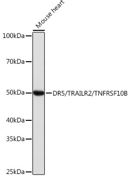
Western blot analysis of extracts of Mouse heart, using DR5/TRAILR2/TNFRSF10B antibody (A19043) at 1:1000 dilution.
Secondary antibody: HRP Goat Anti-Rabbit IgG (H+L) (AS014) at 1:10000 dilution.
Lysates/proteins: 25μg per lane.
Blocking buffer: 3% nonfat dry milk in TBST.
Detection: ECL Basic Kit (RM00020).
Exposure time: 1s.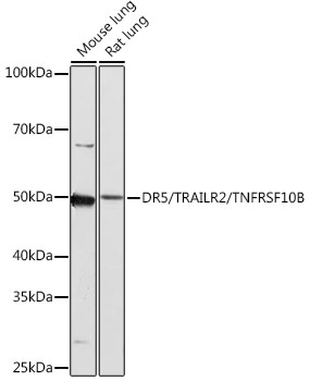
Western blot analysis of extracts of various cell lines, using DR5/TRAILR2/TNFRSF10B antibody (A19043) at 1:1000 dilution.
Secondary antibody: HRP Goat Anti-Rabbit IgG (H+L) (AS014) at 1:10000 dilution.
Lysates/proteins: 25μg per lane.
Blocking buffer: 3% nonfat dry milk in TBST.
Detection: ECL Basic Kit (RM00020).
Exposure time: 3min.
Immunohistochemistry analysis of paraffin-embedded human colon carcinoma using DR5/TRAILR2/TNFRSF10B Rabbit mAb (A19043) at dilution of 1:100 (40x lens).Perform microwave antigen retrieval with 10 mM PBS buffer pH 7.2 before commencing with IHC staining protocol.
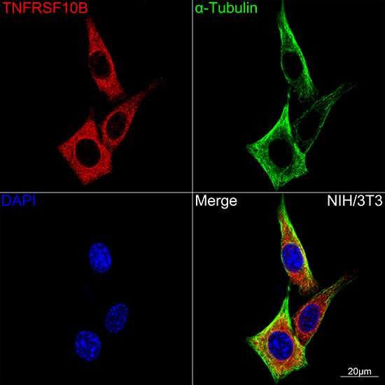
Confocal imaging of NIH-3T3 cells using DR5/TRAILR2/TNFRSF10B Rabbit mAb (A19043, dilution 1:100)(Red). The cells were counterstained with α-Tubulin Mouse mAb (AC012, dilution 1:400) (Green). DAPI was used for nuclear staining (blue). Objective: 100x.
-
Key applications Western blotting, Immunohistochemistry, Immunofluorescence Host species Кролик Antibody type Primary Antibodies Reactivity Человек, Мышь, Крыса Immunogen A synthetic peptide corresponding to a sequence within amino acids 341-440 of human DR5/TRAILR2/TNFRSF10B (O14763). Isotype IgG