


быстрая доставка ~4 недели
Размер
- 100 μL
- 200 μL
- 50 μL
SHP1 Rabbit mAb (A19111)
- Описание
- Характеристики
-
Overview
Product name SHP1 Rabbit mAb Catalog No. A19111 Host species Rabbit Purification method Affinity purification Isotype IgG CloneNo. ARC0295 Background
The protein encoded by this gene is a member of the protein tyrosine phosphatase (PTP) family. PTPs are known to be signaling molecules that regulate a variety of cellular processes including cell growth, differentiation, mitotic cycle, and oncogenic transformation. N-terminal part of this PTP contains two tandem Src homolog (SH2) domains, which act as protein phospho-tyrosine binding domains, and mediate the interaction of this PTP with its substrates. This PTP is expressed primarily in hematopoietic cells, and functions as an important regulator of multiple signaling pathways in hematopoietic cells. This PTP has been shown to interact with, and dephosphorylate a wide spectrum of phospho-proteins involved in hematopoietic cell signaling. Multiple alternatively spliced variants of this gene, which encode distinct isoforms, have been reported.Immunogen information
Immunogen A synthetic peptide corresponding to a sequence within amino acids 496-595 of human SHP1 (P29350). Sequence SGMVQTEAQYKFIYVAIAQFIETTKKKLEVLQSQKGQESEYGNITYPPAMKNAHAKASRTSSKHKEDVYENLHTKNKREEKVKKQRSADKEKSKGSLKRK Gene ID Swiss Prot Synonyms HCP; HCPH; SHP1; SHP-1; HPTP1C; PTP-1C; SHP-1L; SH-PTP1 Calculated MW 68kDa Observed MW 68kDa Applications
Reactivity Human, Mouse, Rat Tested applications WB IHC-P IF/ICC Recommended dilution - WB 1:500 - 1:2000
- IHC-P 1:50 - 1:200
- IF/ICC 1:50 - 1:200
Storage buffer Store at -20℃. Avoid freeze / thaw cycles.
Buffer: PBS with 0.02% sodium azide, 0.05% BSA, 50% glycerol, pH7.3.Key application Western blotting Immunohistochemistry Immunofluorescence Positive samples MCF7, Jurkat, SP2/0, Mouse liver, Rat brain, Raji Cellular location Cytoplasm, Nucleus Customer validation IHC(Homo sapiens)
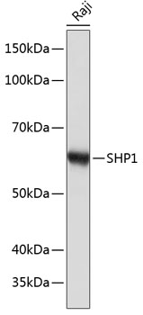
Western blot analysis of extracts of Raji cells, using SHP1 antibody (A19111) at 1:1000 dilution.
Secondary antibody: HRP Goat Anti-Rabbit IgG (H+L) (AS014) at 1:10000 dilution.
Lysates/proteins: 25μg per lane.
Blocking buffer: 3% nonfat dry milk in TBST.
Detection: ECL Basic Kit (RM00020).
Exposure time: 10s.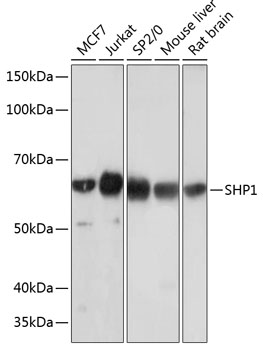
Western blot analysis of extracts of various cell lines, using SHP1 antibody (A19111) at 1:1000 dilution.
Secondary antibody: HRP Goat Anti-Rabbit IgG (H+L) (AS014) at 1:10000 dilution.
Lysates/proteins: 25μg per lane.
Blocking buffer: 3% nonfat dry milk in TBST.
Detection: ECL Basic Kit (RM00020).
Exposure time: 3min.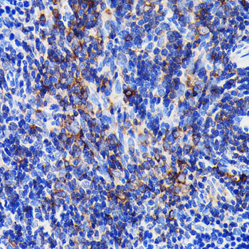
Immunohistochemistry analysis of paraffin-embedded mouse spleen using SHP1 Rabbit mAb (A19111) at dilution of 1:100 (40x lens).Perform microwave antigen retrieval with 10 mM PBS buffer pH 7.2 before commencing with IHC staining protocol.

Immunohistochemistry analysis of paraffin-embedded rat spleen using SHP1 Rabbit mAb (A19111) at dilution of 1:100 (40x lens).Perform microwave antigen retrieval with 10 mM PBS buffer pH 7.2 before commencing with IHC staining protocol.

Immunohistochemistry analysis of paraffin-embedded human appendix using SHP1 Rabbit mAb (A19111) at dilution of 1:100 (40x lens).Perform microwave antigen retrieval with 10 mM PBS buffer pH 7.2 before commencing with IHC staining protocol.

Immunofluorescence analysis of rat spleen using SHP1 Rabbit mAb (A19111) at dilution of 1:100 (40x lens). Blue: DAPI for nuclear staining.
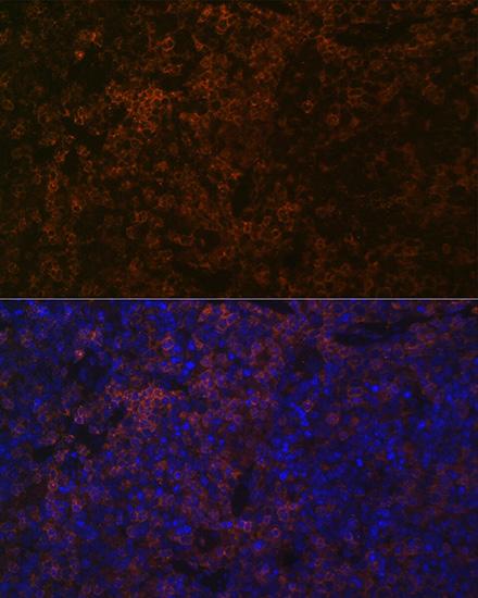
Immunofluorescence analysis of mouse spleen using SHP1 Rabbit mAb (A19111) at dilution of 1:100 (40x lens). Blue: DAPI for nuclear staining.
-
Key applications Western blotting, Immunohistochemistry, Immunofluorescence Host species Кролик Antibody type Primary Antibodies Reactivity Человек, Мышь, Крыса Immunogen A synthetic peptide corresponding to a sequence within amino acids 496-595 of human SHP1 (P29350). Isotype IgG