


быстрая доставка ~4 недели
Размер
- 100 μL
- 200 μL
- 50 μL
PD-L1/CD274 Rabbit mAb (A19135)
- Описание
- Характеристики
-
Overview
Product name PD-L1/CD274 Rabbit mAb Catalog No. A19135 Host species Rabbit Purification method Affinity purification Isotype IgG CloneNo. ARC2478 Background
This gene encodes an immune inhibitory receptor ligand that is expressed by hematopoietic and non-hematopoietic cells, such as T cells and B cells and various types of tumor cells. The encoded protein is a type I transmembrane protein that has immunoglobulin V-like and C-like domains. Interaction of this ligand with its receptor inhibits T-cell activation and cytokine production. During infection or inflammation of normal tissue, this interaction is important for preventing autoimmunity by maintaining homeostasis of the immune response. In tumor microenvironments, this interaction provides an immune escape for tumor cells through cytotoxic T-cell inactivation. Expression of this gene in tumor cells is considered to be prognostic in many types of human malignancies, including colon cancer and renal cell carcinoma. Alternative splicing results in multiple transcript variants.Immunogen information
Immunogen Recombinant fusion protein containing a sequence corresponding to amino acids 1-290 of human PD-L1/CD274 (Q9NZQ7). Sequence MRIFAVFIFMTYWHLLNAFTVTVPKDLYVVEYGSNMTIECKFPVEKQLDLAALIVYWEMEDKNIIQFVHGEEDLKVQHSSYRQRARLLKDQLSLGNAALQITDVKLQDAGVYRCMISYGGADYKRITVKVNAPYNKINQRILVVDPVTSEHELTCQAEGYPKAEVIWTSSDHQVLSGKTTTTNSKREEKLFNVTSTLRINTTTNEIFYCTFRRLDPEENHTAELVIPELPLAHPPNERTHLVILGAILLCLGVALTFIFRLRKGRMMDVKKCGIQDTNSKKQSDTHLEET Gene ID Swiss Prot Synonyms B7-H; B7H1; PDL1; PD-L1; hPD-L1; PDCD1L1; PDCD1LG1 Calculated MW 33kDa Observed MW 40-50kDa Applications
Reactivity Human Tested applications WB IHC-P Recommended dilution - WB 1:500 - 1:1000
- IHC-P 1:100 - 1:500
Storage buffer Store at -20℃. Avoid freeze / thaw cycles.
Buffer: PBS with 0.05% proclin300, 0.05% BSA, 50% glycerol, pH7.3.Key application Western blotting Immunohistochemistry Positive samples A549+IFNG Cellular location Cell membrane, Endomembrane system, Single-pass type I membrane protein 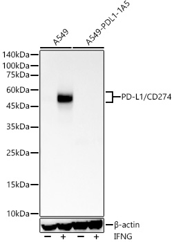
Western blot analysis of lysates from wild type (WT) and PD-L1/CD274 knockout (KO) A549 cells using PD-L1/CD274 Rabbit mAb (A19135) at 1:1000 dilution.wild type (WT) and PD-L1/CD274 knockout (KO) A549 cells were treated by IFNG (100 ng/mL) at 37℃ for 48 hours.
Secondary antibody: HRP Goat Anti-Rabbit IgG (H+L) (AS014) at 1:10000 dilution.
Lysates/proteins: 25ug per lane.
Blocking buffer: 3% nonfat dry milk in TBST.
Detection: ECL Basic Kit (RM00020).
Exposure time: 60s.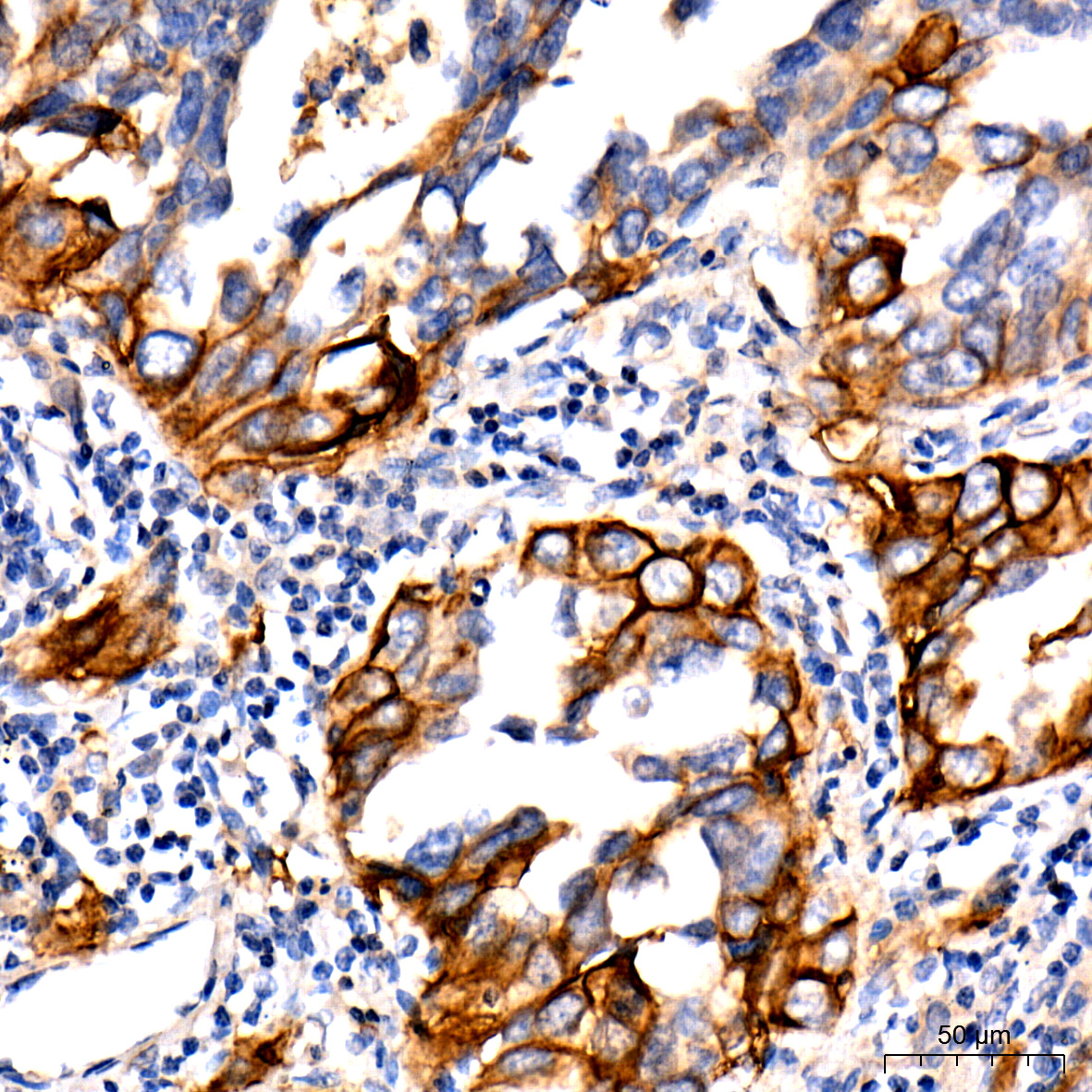
Immunohistochemistry of paraffin-embedded Human lung adenocarcinoma using PD-L1/CD274 Rabbit mAb (A19135) at dilution of 1:500 (40x lens).Perform high pressure antigen retrieval with 10 mM citrate buffer pH 6.0 before commencing with IHC staining protocol.
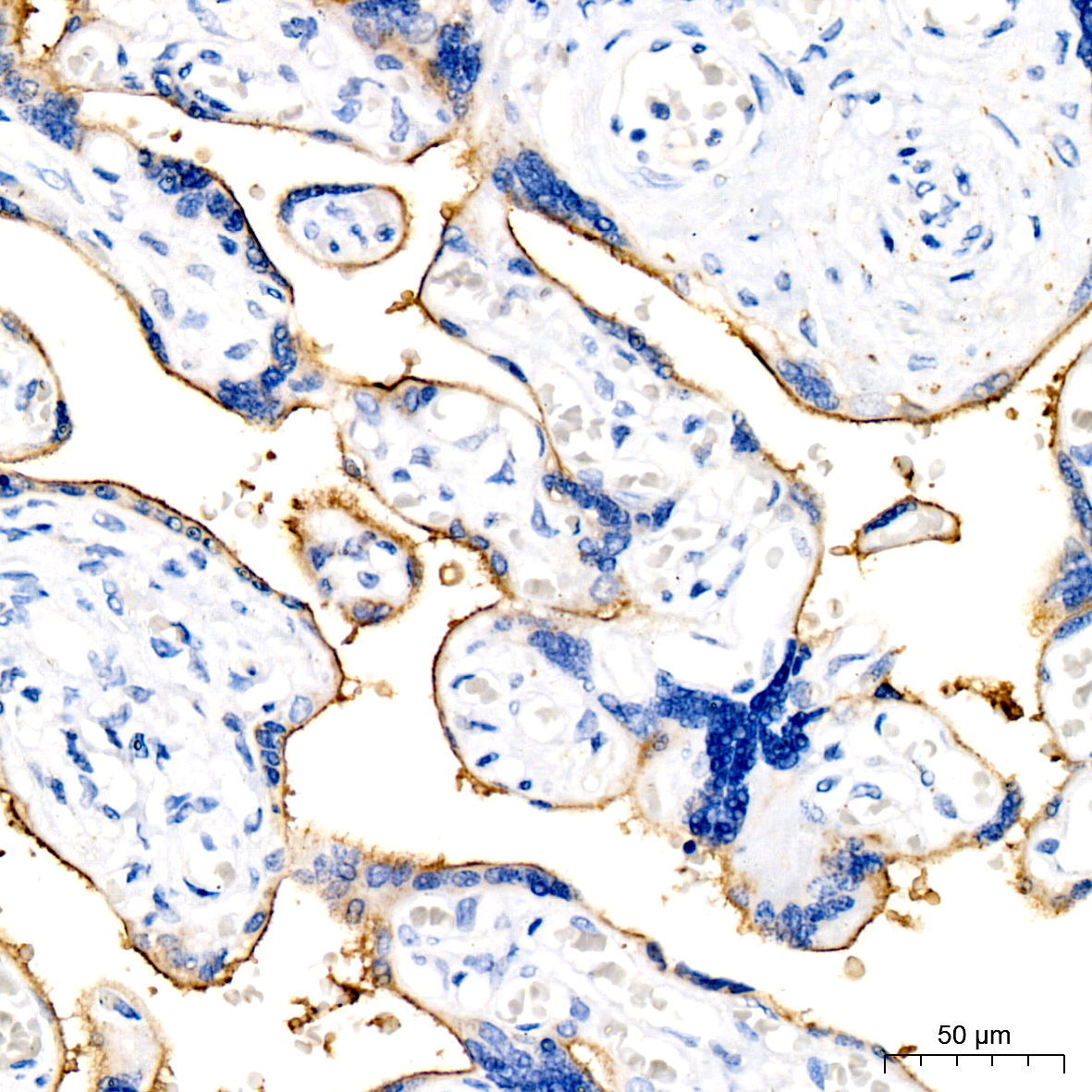
Immunohistochemistry of paraffin-embedded human placenta using PD-L1/CD274 Rabbit mAb (A19135) at dilution of 1:500 (40x lens).Perform high pressure antigen retrieval with 10 mM citrate buffer pH 6.0 before commencing with IHC staining protocol.
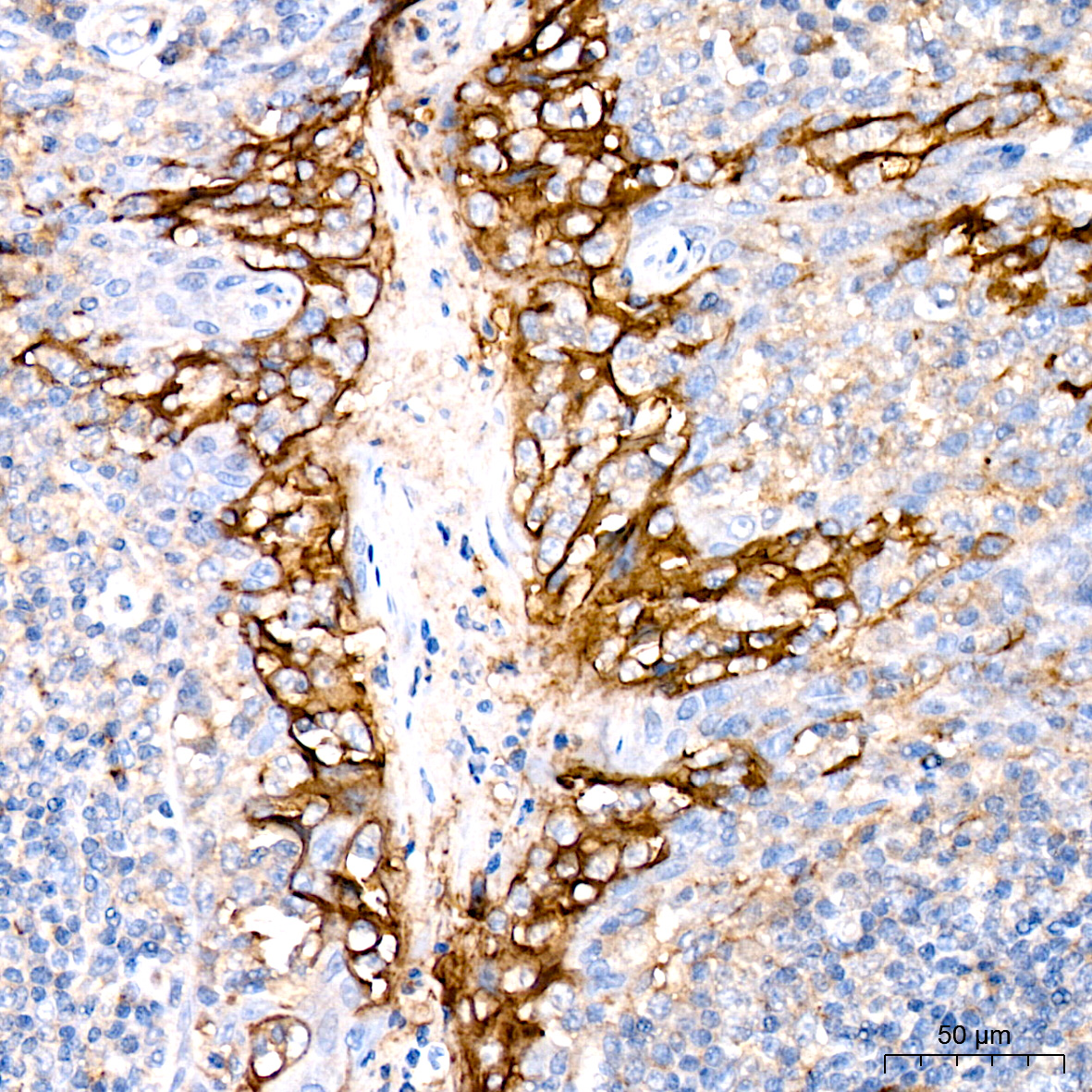
Immunohistochemistry of paraffin-embedded human tonsil using PD-L1/CD274 Rabbit mAb (A19135) at dilution of 1:500 (40x lens).Perform high pressure antigen retrieval with 10 mM citrate buffer pH 6.0 before commencing with IHC staining protocol.
-
Key applications Western blotting, Immunohistochemistry Host species Кролик Antibody type Primary Antibodies Reactivity Человек Immunogen Recombinant fusion protein containing a sequence corresponding to amino acids 1-290 of human PD-L1/CD274 (Q9NZQ7). KO Validated Validated Isotype IgG