


быстрая доставка ~4 недели
Размер
- 100 μL
- 200 μL
- 50 μL
Coagulation factor III/Tissue Factor Rabbit mAb (A23243)
- Описание
- Характеристики
-
Overview
Product name Coagulation factor III/Tissue Factor Rabbit mAb Catalog No. A23243 Host species Rabbit Purification method Affinity purification Isotype IgG CloneNo. ARC60559 Background
This gene encodes coagulation factor III which is a cell surface glycoprotein. This factor enables cells to initiate the blood coagulation cascades, and it functions as the high-affinity receptor for the coagulation factor VII. The resulting complex provides a catalytic event that is responsible for initiation of the coagulation protease cascades by specific limited proteolysis. Unlike the other cofactors of these protease cascades, which circulate as nonfunctional precursors, this factor is a potent initiator that is fully functional when expressed on cell surfaces, for example, on monocytes. There are 3 distinct domains of this factor: extracellular, transmembrane, and cytoplasmic. Platelets and monocytes have been shown to express this coagulation factor under procoagulatory and proinflammatory stimuli, and a major role in HIV-associated coagulopathy has been described. Platelet-dependent monocyte expression of coagulation factor III has been described to be associated with Coronavirus Disease 2019 (COVID-19) severity and mortality. This protein is the only one in the coagulation pathway for which a congenital deficiency has not been described. Alternate splicing results in multiple transcript variants.Immunogen information
Immunogen Recombinant protein of human Coagulation factor III/Tissue Factor. Sequence Email for sequence Gene ID Swiss Prot Synonyms TF; TFA; CD142 Calculated MW 33kDa Observed MW 45-53kDa Applications
Reactivity Human, Mouse Tested applications WB IHC-P IF/ICC Recommended dilution - WB 1:500 - 1:1000
- IHC-P 1:50 - 1:200
- IF/ICC 1:50 - 1:200
Storage buffer Store at -20℃. Avoid freeze / thaw cycles.
Buffer: PBS with 0.05% proclin300, 0.05% BSA, 50% glycerol, pH7.3.Key application Western blotting Immunohistochemistry Immunofluorescence Positive samples BxPC-3, A-431, MDA-MB-231 Cellular location Membrane, Secreted, Single-pass type I membrane protein 
Western blot analysis of various lysates, using Coagulation factor III/Tissue Factor Rabbit mAb (A23243) at 1:1000 dilution.
Secondary antibody: HRP Goat Anti-Rabbit IgG (H+L) (AS014) at 1:10000 dilution.
Lysates/proteins: 25μg per lane.
Blocking buffer: 3% nonfat dry milk in TBST.
Detection: ECL Basic Kit (RM00020).
Exposure time: 10s.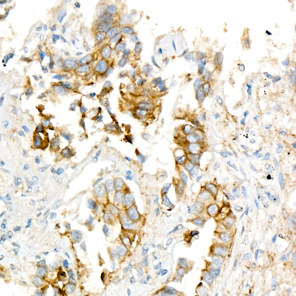
Immunohistochemistry analysis of paraffin-embedded human lung cancer using Coagulation factor III/Tissue Factor Rabbit mAb (A23243) at dilution of 1:400 (40x lens).Perform high pressure antigen retrieval with 10 mM citrate buffer pH 6.0 before commencing with IHC staining protocol.
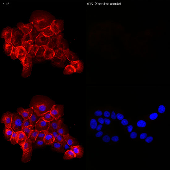
Immunofluorescence analysis of A-431 and MCF7(Negative sample) cells using Coagulation factor III/Tissue Factor Rabbit mAb (A23243) at dilution of 1:300 (40x lens). Blue: DAPI for nuclear staining.
-
Key applications Western blotting, Immunohistochemistry, Immunofluorescence Host species Кролик Antibody type Primary Antibodies Reactivity Человек, Мышь Immunogen Recombinant protein of human Coagulation factor III/Tissue Factor. Isotype IgG