


быстрая доставка ~4 недели
Размер
- 100 μL
- 20 μL
- 200 μL
- 50 μL
DDB1 Rabbit mAb (A5022)
- Описание
- Характеристики
-
Overview
Product name DDB1 Rabbit mAb Catalog No. A5022 Host species Rabbit Purification method Affinity purification Isotype IgG CloneNo. ARC1278 Background
The protein encoded by this gene is the large subunit (p127) of the heterodimeric DNA damage-binding (DDB) complex while another protein (p48) forms the small subunit. This protein complex functions in nucleotide-excision repair and binds to DNA following UV damage. Defective activity of this complex causes the repair defect in patients with xeroderma pigmentosum complementation group E (XPE) - an autosomal recessive disorder characterized by photosensitivity and early onset of carcinomas. However, it remains for mutation analysis to demonstrate whether the defect in XPE patients is in this gene or the gene encoding the small subunit. In addition, Best vitelliform mascular dystrophy is mapped to the same region as this gene on 11q, but no sequence alternations of this gene are demonstrated in Best disease patients. The protein encoded by this gene also functions as an adaptor molecule for the cullin 4 (CUL4) ubiquitin E3 ligase complex by facilitating the binding of substrates to this complex and the ubiquitination of proteins.Immunogen information
Immunogen A synthetic peptide corresponding to a sequence within amino acids 1041-1140 of human DDB1 (NP_001914.3). Sequence TSLSESWYNLLLDMQNRLNKVIKSVGKIEHSFWRSFHTERKTEPATGFIDGDLIESFLDISRPKMQEVVANLQYDDGSGMKREATADDLIKVVEELTRIH Gene ID Swiss Prot Synonyms XPE; DDBA; XAP1; XPCE; XPE-BF; UV-DDB1; WHIKERS Calculated MW 127kDa Observed MW 127kDa Applications
Reactivity Human, Mouse, Rat Tested applications WB IHC-P Recommended dilution - WB 1:500 - 1:2000
- IHC-P 1:50 - 1:200
Storage buffer Store at -20℃. Avoid freeze / thaw cycles.
Buffer: PBS with 0.02% sodium azide, 0.05% BSA, 50% glycerol, pH7.3.Key application Western blotting Immunohistochemistry Positive samples HeLa, MCF7, NIH/3T3, C6, Mouse brain, Rat lung Cellular location Cytoplasm, Nucleus 
Western blot analysis of extracts of various cell lines, using DDB1 Rabbit mAb (A5022) at 1:1000 dilution.
Secondary antibody: HRP Goat Anti-Rabbit IgG (H+L) (AS014) at 1:10000 dilution.
Lysates/proteins: 25μg per lane.
Blocking buffer: 3% nonfat dry milk in TBST.
Detection: ECL Basic Kit (RM00020).
Exposure time: 1s.
Immunohistochemistry analysis of paraffin-embedded rat brain using DDB1 Rabbit mAb (A5022) at dilution of 1:100 (40x lens).Perform microwave antigen retrieval with 10 mM Tris/EDTA buffer pH 9.0 before commencing with IHC staining protocol.
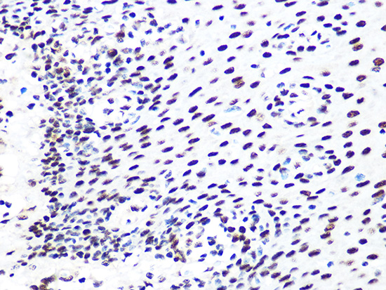
Immunohistochemistry analysis of paraffin-embedded human esophageal using DDB1 Rabbit mAb (A5022) at dilution of 1:100 (40x lens).Perform microwave antigen retrieval with 10 mM Tris/EDTA buffer pH 9.0 before commencing with IHC staining protocol.
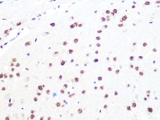
Immunohistochemistry analysis of paraffin-embedded mouse brain using DDB1 Rabbit mAb (A5022) at dilution of 1:100 (40x lens).Perform microwave antigen retrieval with 10 mM Tris/EDTA buffer pH 9.0 before commencing with IHC staining protocol.
-
Key applications Western blotting, Immunohistochemistry Host species Кролик Antibody type Primary Antibodies Reactivity Человек, Мышь, Крыса Immunogen A synthetic peptide corresponding to a sequence within amino acids 1041-1140 of human DDB1 (NP_001914.3). Isotype IgG