


быстрая доставка ~4 недели
Размер
- 100 μL
- 200 μL
- 50 μL
Acetyl-Histone H3-K4 Rabbit pAb (A16078)
- Описание
- Характеристики
-
Overview
Product name Acetyl-Histone H3-K4 Rabbit pAb Catalog No. A16078 Host species Rabbit Purification method Affinity purification Isotype IgG Background
Histones are basic nuclear proteins that are responsible for the nucleosome structure of the chromosomal fiber in eukaryotes. Nucleosomes consist of approximately 146 bp of DNA wrapped around a histone octamer composed of pairs of each of the four core histones (H2A, H2B, H3, and H4). The chromatin fiber is further compacted through the interaction of a linker histone, H1, with the DNA between the nucleosomes to form higher order chromatin structures. This gene is intronless and encodes a replication-dependent histone that is a member of the histone H3 family. Transcripts from this gene lack polyA tails; instead, they contain a palindromic termination element. This gene is located separately from the other H3 genes that are in the histone gene cluster on chromosome 6p22-p21.3.Immunogen information
Immunogen A synthetic acetylated peptide around K4 of human Histone H3 (NP_003484.1). Sequence ARTKQ Gene ID Swiss Prot Synonyms H3t; H3.4; H3/g; H3FT; H3C16; HIST3H3 Calculated MW 16kDa Observed MW 15kDa/17kDa Applications
Reactivity Human, Mouse, Rat, Other (Wide Range) Tested applications WB IF/ICC IP ChIP Recommended dilution - WB 1:500 - 1:1000
- IF/ICC 1:50 - 1:100
- ChIP 1:50 - 1:200
Storage buffer Store at -20℃. Avoid freeze / thaw cycles.
Buffer: PBS with 0.01% thimerosal, 50% glycerol, pH7.3.Key application Western blotting Immunofluorescence Positive samples NIH/3T3, C6 Cellular location Chromosome, Nucleus Customer validation WB(Mus musculus)
IF(Mus musculus)

Western blot analysis of extracts of various cell lines, using Acetyl-Histone H3-K4 antibody (A16078) at 1:1000 dilution.NIH/3T3 cells were treated by TSA (1 uM) at 37℃ for 18 hours.C6 cells were treated by TSA (1 uM) at 37℃ for 18 hours.
Secondary antibody: HRP Goat Anti-Rabbit IgG (H+L) (AS014) at 1:10000 dilution.
Lysates/proteins: 25μg per lane.
Blocking buffer: 3% nonfat dry milk in TBST.
Detection: ECL Basic Kit (RM00020).
Exposure time: 90s.
Western blot analysis of extracts of NIH/3T3 cells, using Acetyl-Histone H3-K4 antibody (A16078) at 1:1000 dilution.NIH/3T3 cells were treated by TSA (1 uM) at 37℃ for 18 hours.
Secondary antibody: HRP Goat Anti-Rabbit IgG (H+L) (AS014) at 1:10000 dilution.
Lysates/proteins: 25μg per lane.
Blocking buffer: 3% nonfat dry milk in TBST.
Detection: ECL Basic Kit (RM00020).
Exposure time: 300s.
Immunofluorescence analysis of C6 cells using Acetyl-Histone H3-K4 antibody (A16078) at dilution of 1:100.C6 cells were treated by TSA (1 uM) at 37℃ for 18 hours. Blue: DAPI for nuclear staining.
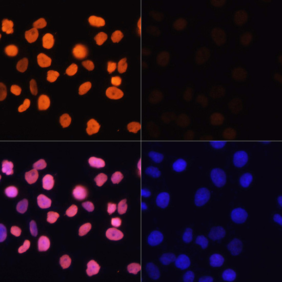
Immunofluorescence analysis of HeLa cells using Acetyl-Histone H3-K4 antibody (A16078) at dilution of 1:100.HeLa cells were treated by TSA (1 uM) at 37℃ for 18 hours. Blue: DAPI for nuclear staining.

Immunofluorescence analysis of NIH/3T3 cells using Acetyl-Histone H3-K4 antibody (A16078) at dilution of 1:100.NIH/3T3 cells were treated by TSA (1 uM) at 37℃ for 18 hours. Blue: DAPI for nuclear staining.
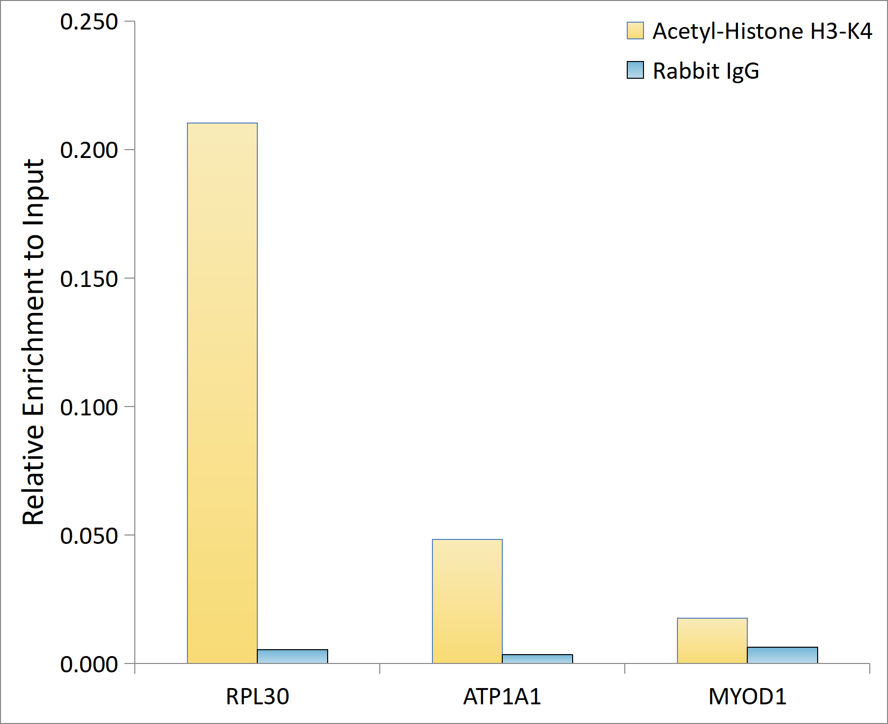
Chromatin immunoprecipitation analysis of extracts of HeLa cells, using Acetyl-Histone H3-K4 antibody (A16078) and rabbit IgG.The amount of immunoprecipitated DNA was checked by quantitative PCR. Histogram was constructed by the ratios of the immunoprecipitated DNA to the input.
-
Key applications Western blotting, Immunofluorescence Host species Кролик Antibody type Primary Antibodies Reactivity Человек, Мышь, Крыса, Другое (Широкий ассортимент) Immunogen A synthetic acetylated peptide around K4 of human Histone H3 (NP_003484.1). Isotype IgG