


![[KO Validated] β-Catenin Rabbit mAb (A19657)](/upload/resize_cache/iblock/3fb/340_340_140cd750bba9870f18aada2478b24840a/e8hejpybrmdfluvvzksucp0imwklj55j.jpg "[KO Validated] β-Catenin Rabbit mAb (A19657)")
быстрая доставка ~4 недели
Размер
- 100 μL
- 200 μL
- 50 μL
[KO Validated] β-Catenin Rabbit mAb (A19657)
- Описание
- Характеристики
-
Overview
Product name [KO Validated] β-Catenin Rabbit mAb Catalog No. A19657 Host species Rabbit Purification method Affinity purification Isotype IgG CloneNo. ARC0136 Background
The protein encoded by this gene is part of a complex of proteins that constitute adherens junctions (AJs). AJs are necessary for the creation and maintenance of epithelial cell layers by regulating cell growth and adhesion between cells. The encoded protein also anchors the actin cytoskeleton and may be responsible for transmitting the contact inhibition signal that causes cells to stop dividing once the epithelial sheet is complete. Finally, this protein binds to the product of the APC gene, which is mutated in adenomatous polyposis of the colon. Mutations in this gene are a cause of colorectal cancer (CRC), pilomatrixoma (PTR), medulloblastoma (MDB), and ovarian cancer. Alternative splicing results in multiple transcript variants.Immunogen information
Immunogen A synthetic peptide corresponding to a sequence within amino acids 1-100 of human beta Catenin (P35222). Sequence MATQADLMELDMAMEPDRKAAVSHWQQQSYLDSGIHSGATTTAPSLSGKGNPEEEDVDTSQVLYEWEQGFSQSFTQEQVADIDGQYAMTRAQRVRAAMFP Gene ID Swiss Prot Synonyms EVR7; CTNNB; MRD19; NEDSDV; armadillo Calculated MW 85kDa Observed MW 92kDa Applications
Reactivity Human, Mouse, Rat Tested applications WB IHC-P IF/ICC IP Recommended dilution - WB 1:500 - 1:1000
- IHC-P 1:50 - 1:200
- IF/ICC 1:50 - 1:200
- IP 1:500 - 1:1000
Storage buffer Store at -20℃. Avoid freeze / thaw cycles.
Buffer: PBS with 0.05% proclin300, 0.05% BSA, 50% glycerol, pH7.3.Key application Western blotting Immunohistochemistry Immunofluorescence Immunoprecipitation Positive samples HeLa cells, 293T cells, NIH/3T3 cells, Mouse brain Cellular location Cell junction, Cell membrane, Cytoplasm, Nucleus, adherens junction, centrosome, cytoskeleton, microtubule organizing center, spindle pole Customer validation (normal mouse hippocampus)
WB(Homo sapiens, Rattus norvegicus, Mus musculus, Mus musculus )
IF(Homo sapiens, Mus musculus)
IHC(Homo sapiens, Mus musculus)
IF(Mus musculus, Homo sapiens)
IF(Homo sapiens, Mus musculus, Rattus norvegicus)
WB(Mus musculus)
Western Blot(Mus musculus)
IHC(Homo sapiens, Rattus norvegicus, Mus musculus)
RT-qPCR(Rattus norvegicus)
Co-IP(Mus musculus)
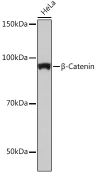
Western blot analysis of extracts of HeLa cells, using β-Catenin antibody (A19657) at 1:1000 dilution.
Secondary antibody: HRP Goat Anti-Rabbit IgG (H+L) (AS014) at 1:10000 dilution.
Lysates/proteins: 25μg per lane.
Blocking buffer: 3% nonfat dry milk in TBST.
Detection: ECL Basic Kit (RM00020).
Exposure time: 10s.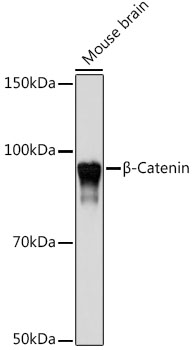
Western blot analysis of extracts of Mouse brain, using β-Catenin antibody (A19657) at 1:1000 dilution.
Secondary antibody: HRP Goat Anti-Rabbit IgG (H+L) (AS014) at 1:10000 dilution.
Lysates/proteins: 25μg per lane.
Blocking buffer: 3% nonfat dry milk in TBST.
Detection: ECL Basic Kit (RM00020).
Exposure time: 10s.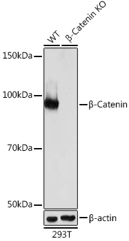
Western blot analysis of extracts from wild type (WT) and β-Catenin knockout (KO) 293T cells, using β-Catenin antibody (A19657) at 1:1000 dilution.
Secondary antibody: HRP Goat Anti-Rabbit IgG (H+L) (AS014) at 1:10000 dilution.
Lysates/proteins: 25μg per lane.
Blocking buffer: 3% nonfat dry milk in TBST.
Detection: ECL Basic Kit (RM00020).
Exposure time: 10s.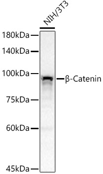
Western blot analysis of NIH/3T3, using β-Catenin antibody (A19657) at 1:1000 dilution.
Lysates/proteins: 25μg per lane.
Blocking buffer: 3% nonfat dry milk in TBST.
Detection: ECL Basic Kit (RM00020).
Exposure time: 30s.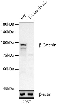
Western blot analysis of extracts from wild type(WT) and [KO Validated] β-Catenin knockout (KO) 293T(KO) cells, using [KO Validated] β-Catenin Rabbit mAb (A19657) at 1:1000 dilution.
Secondary antibody: HRP Goat Anti-Rabbit IgG (H+L) (AS014) at 1:10000 dilution.
Lysates/proteins: 25μg per lane.
Blocking buffer: 3% nonfat dry milk in TBST.
Detection: ECL Basic Kit (RM00020).
Exposure time: 30s.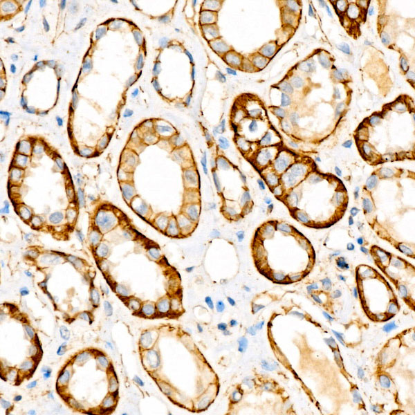
Immunohistochemistry analysis of paraffin-embedded human kidney using [KO Validated] β-Catenin Rabbit mAb (A19657) at dilution of 1:100 (40x lens).Perform high pressure antigen retrieval with 10 mM citrate buffer pH 6.0 before commencing with IHC staining protocol.
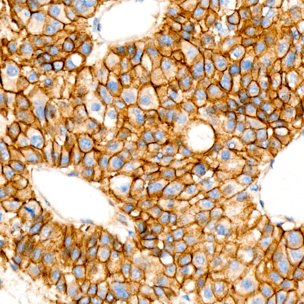
Immunohistochemistry analysis of paraffin-embedded human liver cancer using [KO Validated] β-Catenin Rabbit mAb (A19657) at dilution of 1:100 (40x lens).Perform high pressure antigen retrieval with 10 mM citrate buffer pH 6.0 before commencing with IHC staining protocol.

Immunohistochemistry analysis of paraffin-embedded human liver using [KO Validated] β-Catenin Rabbit mAb (A19657) at dilution of 1:100 (40x lens).Perform high pressure antigen retrieval with 10 mM citrate buffer pH 6.0 before commencing with IHC staining protocol.
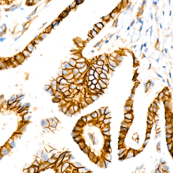
Immunohistochemistry analysis of paraffin-embedded human thyroid cancer using [KO Validated] β-Catenin Rabbit mAb (A19657) at dilution of 1:100 (40x lens).Perform high pressure antigen retrieval with 10 mM citrate buffer pH 6.0 before commencing with IHC staining protocol.

Immunofluorescence analysis of C6 using [KO Validated] β-Catenin Rabbit mAb (A19657) at dilution of 100 (40x lens). Blue: DAPI for nuclear staining.
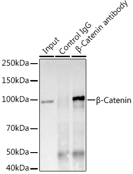
Immunoprecipitation analysis of 600 μg extracts of Mouse brain using 3 μg β-Catenin antibody (A19657). Western blot was performed from the immunoprecipitate using β-Catenin (A19657) at a dilution of 1:1000.
-
Key applications Western blotting, Immunohistochemistry, Immunofluorescence, Immunoprecipitation Host species Кролик Antibody type Primary Antibodies Reactivity Человек, Мышь, Крыса Immunogen A synthetic peptide corresponding to a sequence within amino acids 1-100 of human beta Catenin (P35222). KO Validated Validated Isotype IgG