


![[KO Validated] TTC11/FIS1 Rabbit mAb (A19666)](/upload/resize_cache/iblock/bd6/340_340_140cd750bba9870f18aada2478b24840a/upg4l8v763kay9taqfyaruc33hlub9sa.jpg "[KO Validated] TTC11/FIS1 Rabbit mAb (A19666)")
быстрая доставка ~4 недели
Размер
- 100 μL
- 200 μL
- 50 μL
[KO Validated] TTC11/FIS1 Rabbit mAb (A19666)
- Описание
- Характеристики
-
Overview
Product name [KO Validated] TTC11/FIS1 Rabbit mAb Catalog No. A19666 Host species Rabbit Purification method Affinity purification Isotype IgG CloneNo. ARC5010-03 Background
Enables identical protein binding activity. Involved in several processes, including calcium-mediated signaling using intracellular calcium source; cellular calcium ion homeostasis; and mitochondrion organization. Acts upstream of or within mitochondrion morphogenesis. Located in mitochondrion and peroxisome. Is integral component of mitochondrial outer membrane and integral component of peroxisomal membrane. Part of protein-containing complex. Biomarker of Alzheimer's disease.Immunogen information
Immunogen Recombinant fusion protein containing a sequence corresponding to amino acids 1-122 of human TTC11/FIS1 (NP_057152.2). Sequence MEAVLNELVSVEDLLKFEKKFQSEKAAGSVSKSTQFEYAWCLVRSKYNDDIRKGIVLLEELLPKGSKEEQRDYVFYLAVGNYRLKEYEKALKYVRGLLQTEPQNNQAKELERLIDKAMKKDG Gene ID Swiss Prot Synonyms TTC11; CGI-135 Calculated MW 17kDa Observed MW 15kDa Applications
Reactivity Human, Mouse, Rat Tested applications WB IHC-P IF/ICC IP Recommended dilution - WB 1:500 - 1:1000
- IHC-P 1:50 - 1:200
- IF/ICC 1:50 - 1:200
- IP 1:1000 - 1:5000
Storage buffer Store at -20℃. Avoid freeze / thaw cycles.
Buffer: PBS with 0.01% thimerosal, 0.05% BSA, 50% glycerol, pH7.3.Key application Western blotting Immunohistochemistry Immunofluorescence Immunoprecipitation Positive samples HeLa, HepG2, Mouse heart, Rat spleen, Rat brain Cellular location Mitochondrion outer membrane, Peroxisome membrane, Single-pass membrane protein Customer validation WB(Mus musculus, Rattus norvegicus, Sus scrofa)
IHC(Mus musculus,Rattus norvegicus)
IF(Mus musculus,Rattus norvegicus)
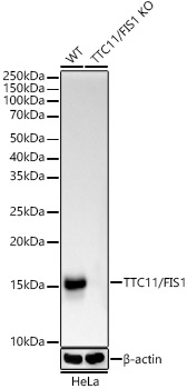
Western blot analysis of extracts from normal (control) and TTC11/FIS1 knockout (KO) HeLa(KO) cells, using TTC11/FIS1 antibody (A19666) at 1:1000 dilution.
Secondary antibody: HRP Goat Anti-Rabbit IgG (H+L) (AS014) at 1:10000 dilution.
Lysates/proteins: 25μg per lane.
Blocking buffer: 3% nonfat dry milk in TBST.
Detection: ECL Basic Kit (RM00020).
Exposure time: 10s.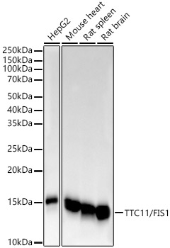
Western blot analysis of various lysates, using TTC11/FIS1 antibody (A19666) at 1:1000 dilution.
Secondary antibody: HRP Goat Anti-Rabbit IgG (H+L) (AS014) at 1:10000 dilution.
Lysates/proteins: 25μg per lane.
Blocking buffer: 3% nonfat dry milk in TBST.
Detection: ECL Basic Kit (RM00020).
Exposure time: 10s.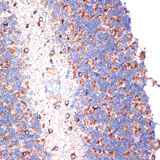
Immunohistochemistry analysis of paraffin-embedded rat brain using [KO Validated] TTC11/FIS1 Rabbit mAb (A19666) at dilution of 1:100 (40x lens).Perform microwave antigen retrieval with 10 mM Tris/EDTA buffer pH 9.0 before commencing with IHC staining protocol.
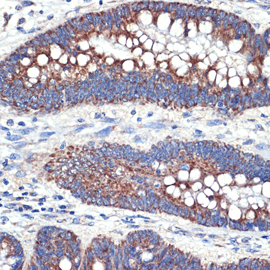
Immunohistochemistry analysis of paraffin-embedded human colon carcinoma using [KO Validated] TTC11/FIS1 Rabbit mAb (A19666) at dilution of 1:100 (40x lens).Perform microwave antigen retrieval with 10 mM Tris/EDTA buffer pH 9.0 before commencing with IHC staining protocol.
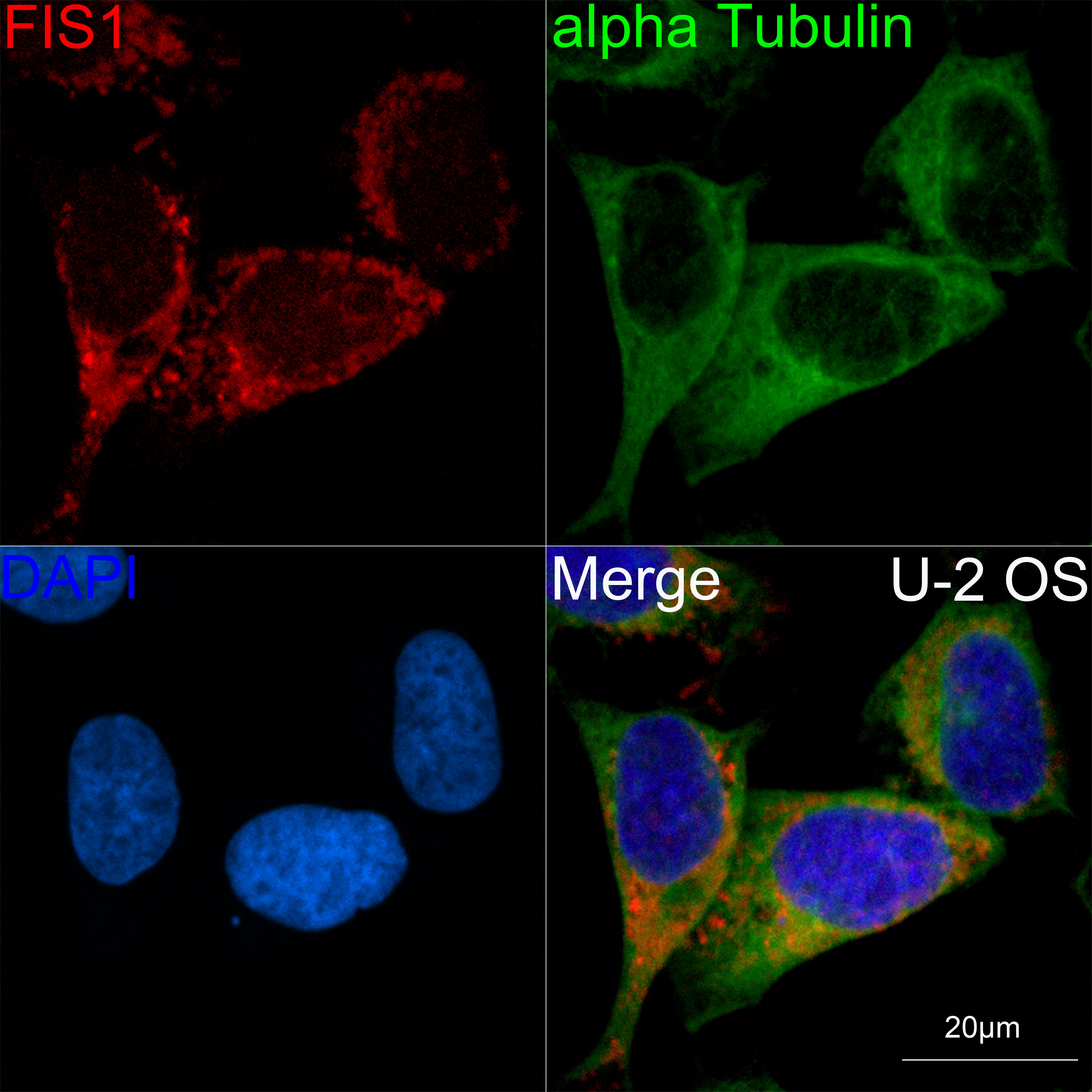
Confocal imaging of U-2 OS cells using [KO Validated] TTC11/FIS1 Rabbit mAb (A19666, dilution 1:100)(Red). The cells were counterstained with α-Tubulin Mouse mAb (AC012, dilution 1:400) (Green). DAPI was used for nuclear staining (blue). Objective: 60x.
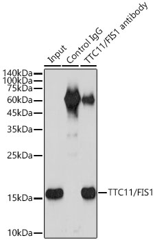
Immunoprecipitation analysis of 300 μg extracts of HeLa cells using 3 μg TTC11/FIS1 antibody (A19666). Western blot was performed from the immunoprecipitate using TTC11/FIS1 (A19666) at a dilution of 1:2000.
-
Key applications Western blotting, Immunohistochemistry, Immunofluorescence, Immunoprecipitation Host species Кролик Antibody type Primary Antibodies Reactivity Человек, Мышь, Крыса Immunogen Recombinant fusion protein containing a sequence corresponding to amino acids 1-122 of human TTC11/FIS1 (NP_057152.2). KO Validated Validated Isotype IgG