


![[KO Validated] CDK9 Rabbit mAb (A11145)](/upload/resize_cache/iblock/ffe/340_340_140cd750bba9870f18aada2478b24840a/hwctzdw553h14hwygvlvgyo1bko456j9.jpg "[KO Validated] CDK9 Rabbit mAb (A11145)")
быстрая доставка ~4 недели
Размер
- 100 μL
- 200 μL
- 50 μL
[KO Validated] CDK9 Rabbit mAb (A11145)
- Описание
- Характеристики
-
Overview
Product name [KO Validated] CDK9 Rabbit mAb Catalog No. A11145 Host species Rabbit Purification method Affinity purification Isotype IgG CloneNo. ARC0527 Background
The protein encoded by this gene is a member of the cyclin-dependent protein kinase (CDK) family. CDK family members are highly similar to the gene products of S. cerevisiae cdc28, and S. pombe cdc2, and known as important cell cycle regulators. This kinase was found to be a component of the multiprotein complex TAK/P-TEFb, which is an elongation factor for RNA polymerase II-directed transcription and functions by phosphorylating the C-terminal domain of the largest subunit of RNA polymerase II. This protein forms a complex with and is regulated by its regulatory subunit cyclin T or cyclin K. HIV-1 Tat protein was found to interact with this protein and cyclin T, which suggested a possible involvement of this protein in AIDS.Immunogen information
Immunogen A synthetic peptide corresponding to a sequence within amino acids 273-372 of human CDK9 (P50750). Sequence RKVKDRLKAYVRDPYALDLIDKLLVLDPAQRIDSDDALNHDFFWSDPMPSDLKGMLSTHLTSMFEYLAPPRRKGSQITQQSTNQSRNPATTNQTEFERVF Gene ID Swiss Prot Synonyms TAK; C-2k; CTK1; CDC2L4; PITALRE Calculated MW 43kDa Observed MW 42kDa/55kDa Applications
Reactivity Human, Mouse, Rat Tested applications WB IHC-P IF/ICC IP ChIP ChIP-seq CUT&Tag Recommended dilution - WB 1:100 - 1:500
- IHC-P 1:50 - 1:200
- IF/ICC 1:50 - 1:200
- IP 1:500 - 1:1000
- ChIP 1:50 - 1:200
- ChIP-seq 2-4μg/IP
- CUT&Tag 10⁵ cells /1 μg
Storage buffer Store at -20℃. Avoid freeze / thaw cycles.
Buffer: PBS with 0.02% sodium azide, 0.05% BSA, 50% glycerol, pH7.3.Key application Western blotting Immunohistochemistry Immunofluorescence Immunoprecipitation Positive samples HeLa, 293T , MCF7, Mouse brain, Rat testis Cellular location Cytoplasm, Nucleus, PML body Customer validation WB(Homo sapiens)
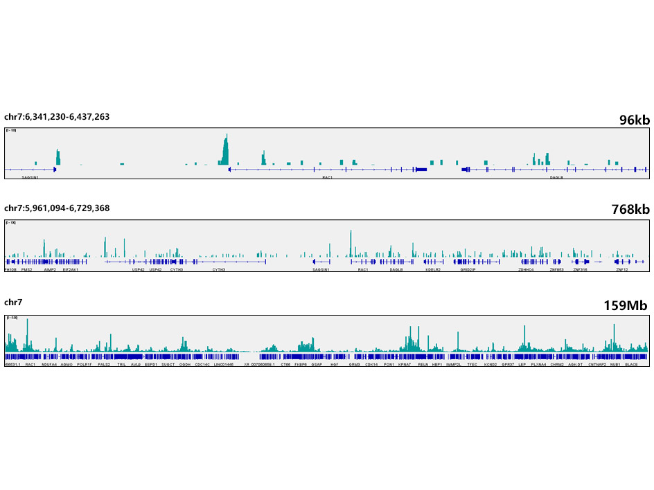
CUT&Tag was performed using the CUT&Tag Assay Kit (pAG-Tn5) for Illumina(RK20265) from 10⁵ K562 cells with 1 μg CDK9 Rabbit mAb (A11145), along with a Goat Anti-Rabbit IgG(H+L). The CUT&Tag results indicate the enrichment pattern of CDK9 in representative gene loci (RAC1), as shown in figure.

Western blot analysis of extracts of various cell lines, using CDK9 Rabbit mAb (A11145) at 1:1000 dilution.
Secondary antibody: HRP Goat Anti-Rabbit IgG (H+L) (AS014) at 1:10000 dilution.
Lysates/proteins: 25μg per lane.
Blocking buffer: 3% nonfat dry milk in TBST.
Detection: ECL Basic Kit (RM00020).
Exposure time: 3min.
Western blot analysis of extracts from wild type(WT) and CDK9 knockout (KO) 293T cells, using CDK9 antibody (A11145) at 1:500 dilution.
Secondary antibody: HRP Goat Anti-Rabbit IgG (H+L) (AS014) at 1:10000 dilution.
Lysates/proteins: 25μg per lane.
Blocking buffer: 3% nonfat dry milk in TBST.
Detection: ECL Basic Kit (RM00020).
Exposure time: 1s.
Immunohistochemistry of paraffin-embedded human colon carcinoma using [KO Validated] CDK9 Rabbit mAb (A11145) at dilution of 1:200 (40x lens).Perform high pressure antigen retrieval with 10 mM citrate buffer pH 6.0 before commencing with IHC staining protocol.

Immunohistochemistry of paraffin-embedded human liver cancer using [KO Validated] CDK9 Rabbit mAb (A11145) at dilution of 1:200 (40x lens).Perform high pressure antigen retrieval with 10 mM citrate buffer pH 6.0 before commencing with IHC staining protocol.

Immunohistochemistry of paraffin-embedded mouse brain using [KO Validated] CDK9 Rabbit mAb (A11145) at dilution of 1:200 (40x lens).Perform high pressure antigen retrieval with 10 mM citrate buffer pH 6.0 before commencing with IHC staining protocol.
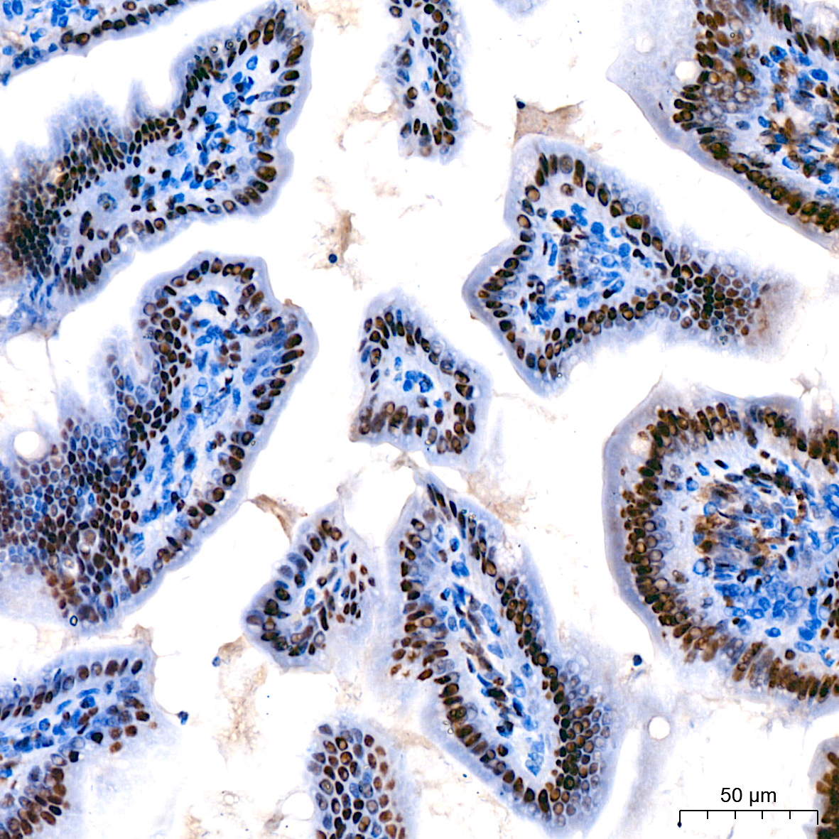
Immunohistochemistry of paraffin-embedded mouse colon using [KO Validated] CDK9 Rabbit mAb (A11145) at dilution of 1:200 (40x lens).Perform high pressure antigen retrieval with 10 mM citrate buffer pH 6.0 before commencing with IHC staining protocol.

Immunohistochemistry of paraffin-embedded mouse liver using [KO Validated] CDK9 Rabbit mAb (A11145) at dilution of 1:200 (40x lens).Perform high pressure antigen retrieval with 10 mM citrate buffer pH 6.0 before commencing with IHC staining protocol.

Immunohistochemistry of paraffin-embedded rat kidney using [KO Validated] CDK9 Rabbit mAb (A11145) at dilution of 1:200 (40x lens).Perform high pressure antigen retrieval with 10 mM citrate buffer pH 6.0 before commencing with IHC staining protocol.

Confocal imaging of U-2 0S cells using [KO Validated] CDK9 Rabbit mAb (A11145, dilution 1:50)(Red). The cells were counterstained with α-Tubulin Mouse mAb (AC012, dilution 1:400) (Green). DAPI was used for nuclear staining (blue). Objective: 60x.

Immunoprecipitation analysis of 200 μg extracts of HeLa cells using 3 μg CDK9 antibody (A11145). Western blot was performed from the immunoprecipitate using CDK9 antibody (A11145) at a dilution of 1:1000.

Chromatin immunoprecipitations were performed with cross-linked chromatin from DLD-1 cells and CDK9 Rabbit mAb (A11145). The ChIP sequencing results indicate the enrichment pattern of CDK9 in selected genomic region and representative gene loci (e.g. RAC1, GAPDH and ACTB), as shown in figure.
-
Key applications Western blotting, Immunohistochemistry, Immunofluorescence, Immunoprecipitation Host species Кролик Antibody type Primary Antibodies Reactivity Человек, Мышь, Крыса Immunogen A synthetic peptide corresponding to a sequence within amino acids 273-372 of human CDK9 (P50750). KO Validated Validated Isotype IgG