


![[KO Validated] AMPKβ1 Rabbit pAb (A12491)](/upload/resize_cache/iblock/dc1/340_340_140cd750bba9870f18aada2478b24840a/yvfjqnj589fgc37dnrx51g115y7u8yku.jpg "[KO Validated] AMPKβ1 Rabbit pAb (A12491)")
быстрая доставка ~4 недели
Размер
- 100 μL
- 200 μL
- 50 μL
[KO Validated] AMPKβ1 Rabbit pAb (A12491)
- Описание
- Характеристики
-
Overview
Product name [KO Validated] AMPKβ1 Rabbit pAb Catalog No. A12491 Host species Rabbit Purification method Affinity purification Isotype IgG Background
The protein encoded by this gene is a regulatory subunit of the AMP-activated protein kinase (AMPK). AMPK is a heterotrimer consisting of an alpha catalytic subunit, and non-catalytic beta and gamma subunits. AMPK is an important energy-sensing enzyme that monitors cellular energy status. In response to cellular metabolic stresses, AMPK is activated, and thus phosphorylates and inactivates acetyl-CoA carboxylase (ACC) and beta-hydroxy beta-methylglutaryl-CoA reductase (HMGCR), key enzymes involved in regulating de novo biosynthesis of fatty acid and cholesterol. This subunit may be a positive regulator of AMPK activity. The myristoylation and phosphorylation of this subunit have been shown to affect the enzyme activity and cellular localization of AMPK. This subunit may also serve as an adaptor molecule mediating the association of the AMPK complex.Immunogen information
Immunogen Recombinant fusion protein containing a sequence corresponding to amino acids 1-80 of human AMPKβ1 (NP_006244.2). Sequence MGNTSSERAALERHGGHKTPRRDSSGGTKDGDRPKILMDSPEDADLFHSEEIKAPEKEEFLAWQHDLEVNDKAPAQARPT Gene ID Swiss Prot Synonyms AMPK; HAMPKb Calculated MW 30kDa Observed MW 38kDa Applications
Reactivity Human, Mouse, Rat Tested applications WB IF/ICC Recommended dilution - WB 1:500 - 1:2000
- IF/ICC 1:50 - 1:100
Storage buffer Store at -20℃. Avoid freeze / thaw cycles.
Buffer: PBS with 0.02% sodium azide, 50% glycerol, pH7.3.Key application Western blotting Immunofluorescence Positive samples U-87MG, A-549, HeLa, Jurkat, HepG2 Cellular location cytoplasm, cytosol, nucleoplasm, nucleus Customer validation WB(Mus musculus)
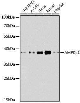
Western blot analysis of extracts of various cell lines, using AMPKβ1 antibody (A12491) at 1:1000 dilution.
Secondary antibody: HRP Goat Anti-Rabbit IgG (H+L) (AS014) at 1:10000 dilution.
Lysates/proteins: 25μg per lane.
Blocking buffer: 3% nonfat dry milk in TBST.
Detection: ECL Basic Kit (RM00020).
Exposure time: 90s.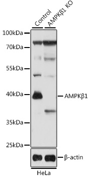
Western blot analysis of extracts from normal (control) and AMPKβ1 knockout (KO) HeLa cells, using AMPKβ1 antibody (A12491) at 1:1000 dilution.
Secondary antibody: HRP Goat Anti-Rabbit IgG (H+L) (AS014) at 1:10000 dilution.
Lysates/proteins: 25μg per lane.
Blocking buffer: 3% nonfat dry milk in TBST.
Detection: ECL Basic Kit (RM00020).
Exposure time: 5s.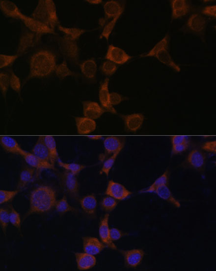
Immunofluorescence analysis of NIH-3T3 cells using AMPKβ1 Polyclonal Antibody (A12491) at dilution of 1:100 (40x lens). Blue: DAPI for nuclear staining.
-
Key applications Western blotting, Immunofluorescence Host species Кролик Antibody type Primary Antibodies Reactivity Человек, Мышь, Крыса Immunogen Recombinant fusion protein containing a sequence corresponding to amino acids 1-80 of human AMPKβ1 (NP_006244.2). KO Validated Validated Isotype IgG