


быстрая доставка ~4 недели
Размер
- 100 μL
- 20 μL
- 200 μL
- 50 μL
Phospho-AKT1-T308+AKT2-T309+AKT3-T305 Rabbit pAb (AP1266)
- Описание
- Характеристики
-
Overview
Product name Phospho-AKT1-T308+AKT2-T309+AKT3-T305 Rabbit pAb Catalog No. AP1266 Host species Rabbit Purification method Affinity purification Isotype IgG Background
The serine-threonine protein kinase encoded by the AKT1 gene is catalytically inactive in serum-starved primary and immortalized fibroblasts. AKT1 and the related AKT2 are activated by platelet-derived growth factor. The activation is rapid and specific, and it is abrogated by mutations in the pleckstrin homology domain of AKT1. It was shown that the activation occurs through phosphatidylinositol 3-kinase. In the developing nervous system AKT is a critical mediator of growth factor-induced neuronal survival. Survival factors can suppress apoptosis in a transcription-independent manner by activating the serine/threonine kinase AKT1, which then phosphorylates and inactivates components of the apoptotic machinery. Mutations in this gene have been associated with the Proteus syndrome. Multiple alternatively spliced transcript variants have been found for this gene. [provided by RefSeq, Jul 2011]Immunogen information
Immunogen A synthetic phosphorylated peptide around T308 of human AktAKT1 (NP_005154.2). Sequence MKTFC Gene ID Swiss Prot Synonyms AKT1/AKT2/AKT3 Calculated MW 48kDa/55kDa/51kDa/54kDa Observed MW 60kDa Applications
Reactivity Human, Mouse, Rat Tested applications WB Recommended dilution - WB 1:500 - 1:1000
Storage buffer Store at -20℃. Avoid freeze / thaw cycles.
Buffer: PBS with 0.05% proclin300, 50% glycerol, pH7.3.Key application Western blotting Positive samples Jurkat Cellular location ciliary basal body, cytoplasm, cytosol, microtubule cytoskeleton, mitochondrion, nucleoplasm, nucleus, plasma membrane, spindle 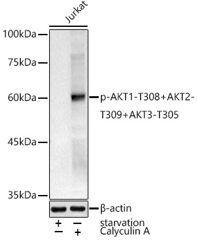
Western blot analysis of extracts of Jurkat cells, using Phospho-AKT1-T308+AKT2-T309+AKT3-T305 antibody (AP1266) at 1:1000 dilution.Jurkat cells were treated by Serum-starvation overnight at 37℃.Jurkat cells were treated by Calyculin A (100 nM) at 37℃ for 30 minutes after serum-starvation overnight.
Secondary antibody: HRP Goat Anti-Rabbit IgG (H+L) (AS014) at 1:10000 dilution.
Lysates/proteins: 25μg per lane.
Blocking buffer: 3% nonfat dry milk in TBST.
Detection: ECL Basic Kit (RM00020).
Exposure time: 180s. -
Key applications Western blotting Host species Кролик Antibody type Primary Antibodies Reactivity Человек, Мышь, Крыса Immunogen A synthetic phosphorylated peptide around T308 of human AktAKT1 (NP_005154.2). Isotype IgG